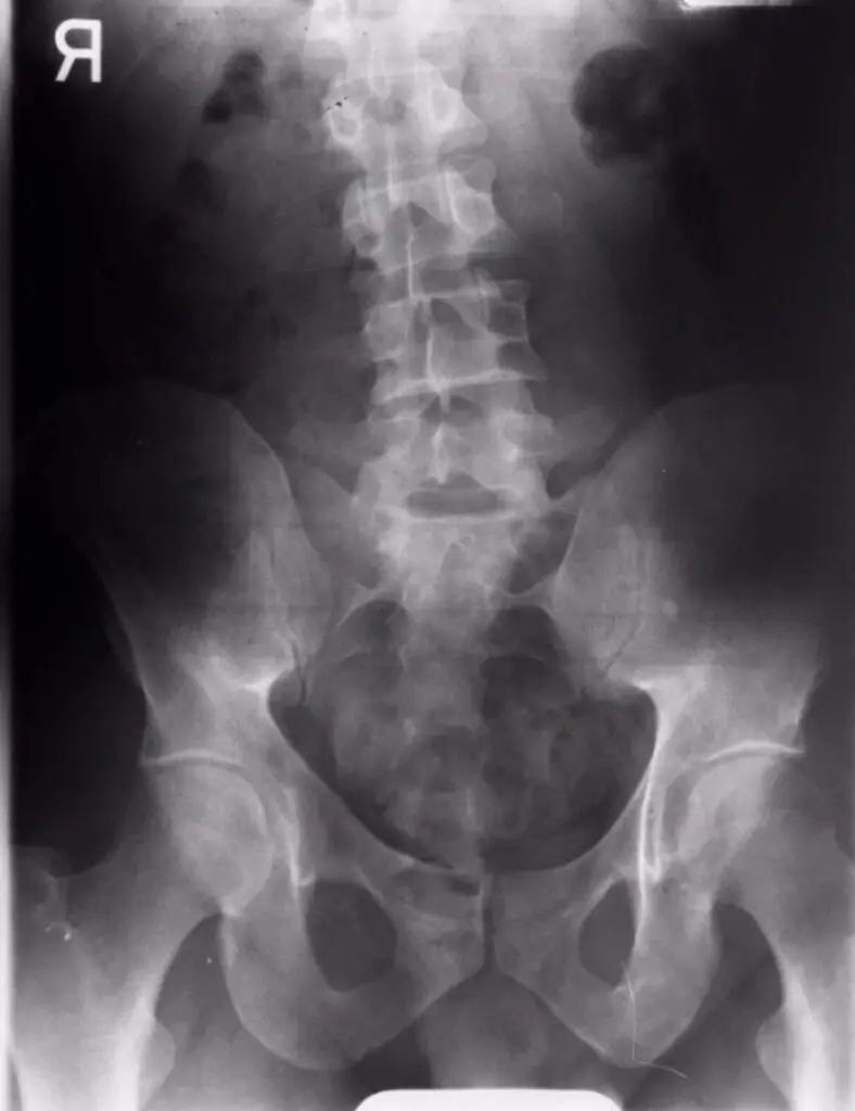
Chiropractic care has evolved considerably from traditional practices such as routine X-rays. It’s essential to comprehend why this change is necessary and what the most recent scientific evidence advises.
Previously, it was commonplace for chiropractic patients to have an X-ray before receiving care. Modern research, however, discourages routine X-rays, indicating they can foster detrimental beliefs about a patient’s pain. The potential harm of unnecessary imaging was underscored in a 2017 British Journal of Sports Medicine editorial titled “It is time to stop causing harm with inappropriate imaging for low back pain.”
Understanding the Risks: How Imaging Can Cause Harm
Unwarranted imaging could be harmful in three primary ways:
- Misinterpretation by Clinicians: Erroneous advice can result from misreading reports, leading to unnecessary investigations and, potentially, invasive procedures like surgery.
- Misinterpretation by Patients: Misunderstood reports can result in fear, catastrophizing, avoidance of activity, and low recovery expectations.
- Imaging Side Effects: One significant side effect is radiation exposure.
Though imaging may offer a detailed view of one’s spinal structures, ‘abnormal findings’ are common in asymptomatic individuals, particularly as they age, and don’t necessarily correlate with pain or disability levels. Moreover, interpretations of images can incite negative beliefs about spine vulnerability, leading to fear and subsequent protective behaviours. Therefore, one must balance the possible benefits and risks of imaging.
Decoding the Latest Evidence
A 2018 paper, “Current Evidence for Spinal X-ray Use in the Chiropractic Profession: a narrative review,” cautioned against routine imaging. It asserted that, in most chiropractic cases, potential harm outweighs any possible benefit from spinal X-rays. Consequently, X-rays shouldn’t be a standard procedure in chiropractic care. Instead, evidence-based clinical guidelines and clinician discretion should dictate the use of diagnostic imaging.
Australian researcher Hazel Jenkins echoed these findings in 2021: “Diagnostic imaging did not result in better clinical outcomes in patients with low back pain presenting for chiropractic care. These results support that current guideline recommendations against routine imaging apply equally to chiropractic practice”.
So, When is Imaging Needed?
You chiropractor should embrace an evidence-based approach, opting for imaging only when necessary. We might order an MRI or CT scan in cases of progressive muscle weakness or condition worsening over time, as these tests offer greater detail of discs and other soft tissues. X-rays might be necessary if we suspect a compression, coccyx fracture, or degenerating hip. In the case of unresponsive disc herniation, we may resort to an MRI or CT scan but not an X-ray. Therefore, the decision to conduct diagnostic imaging should be patient-specific and guided by clinicians’ professional judgment. When is imaging necessary?


