Our patients often ask what causes low back pain. Low back pain can stem from various sources within the spine and surrounding structures. Understanding these primary sources is crucial for effective diagnosis and treatment. Here are the proven primary sources of low back pain.
Intervertebral Disc Issues
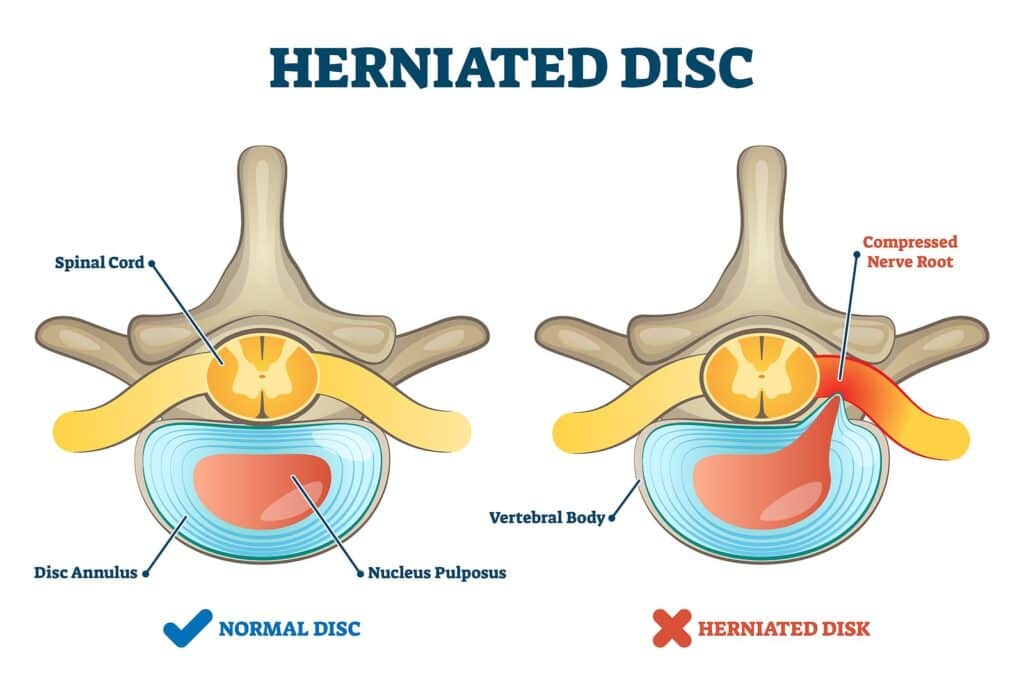
- Herniated Discs: When the inner gel-like core of a disc protrudes through its outer layer, it can compress nearby nerves, causing pain, numbness, and weakness.
- Degenerative Disc Disease: Discs can lose their hydration and elasticity over time, leading to reduced cushioning and increased pain from loading facet joints.
Facet Joint Dysfunction
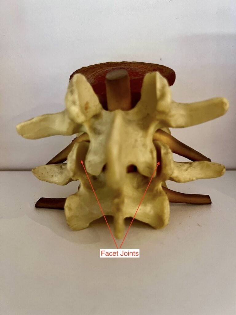
- Facet Joint Syndrome: These small joints between vertebrae can become arthritic or inflamed, leading to localised back pain and stiffness.
- Facet Joint Impingement: Misalignment or locking of these joints can cause acute pain and restricted movement.
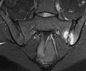
Sacroiliac (SI) Joint Dysfunction
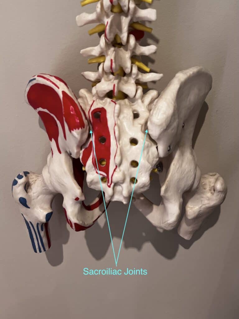
- Sacroiliac Joint Dysfunction: Abnormal movement or alignment of the SI joints, often due to trauma, pregnancy, or arthritis, can cause significant lower back pain.
- Sacroiliitis: Inflammation of the SI joints, often related to arthritis, can result in chronic pain.
Muscle and Ligament Strains
- Muscle Strains: Overexertion, improper lifting, or sudden movements can strain back muscles, leading to pain and spasms. These are usually short-lived, but muscles can become chronically sore when overworked to protect a dysfunctional disc or joint.
- Ligament Sprains: Similar activities can stretch or tear the ligaments in the back, resulting in pain and limited mobility. Ligamentous tears are usually associated with car or sporting accidents.
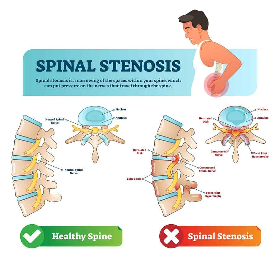
Spinal Stenosis
Narrowing of the Spinal Canal: This condition, often due to ageing, can compress the spinal cord and nerves, causing pain, numbness, and weakness, especially during walking or standing.
Structures like the ligamentum flavum can thicken, leaving less room for the spinal cord and nerves.
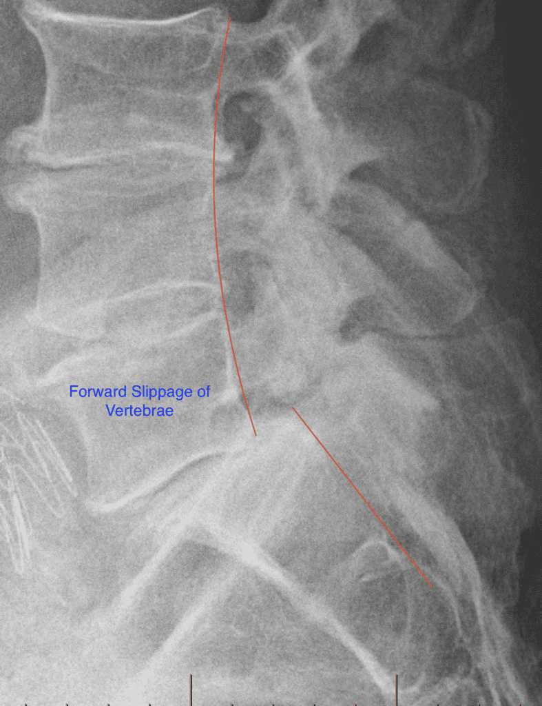
Spondylolisthesis
Vertebral Slippage: When a vertebra slips out of place, usually due to stress fractures in childhood or teenage years, it can compress nerves and cause lower back pain, leg pain and weakness.
Osteoarthritis
Degeneration of Joint Cartilage: This common form of arthritis can affect the spine, leading to pain, stiffness, and reduced flexibility.
It is important to note that the presence of osteoarthritis on an X-ray does not necessarily mean it is causing pain. Many asymptomatic people have osteoarthritis and no pain.
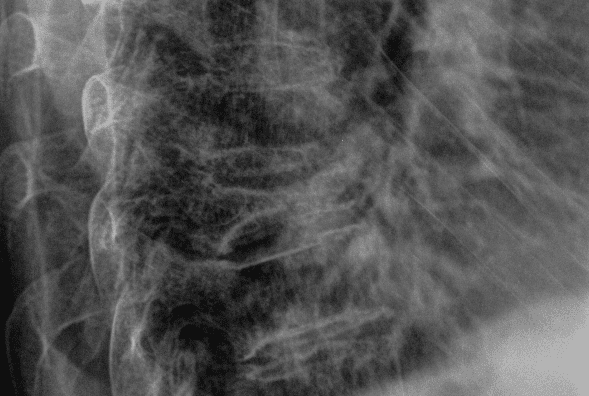
Compression Fractures
Fractured Vertebrae: Often due to osteoporosis, these fractures can cause sudden, severe back pain and decreased spinal height. These are usually seen in osteoporotic patients but can affect people when there has been compressive trauma.
Inflammatory Conditions
Ankylosing Spondylitis: This inflammatory disease primarily affects the spine and pelvis and can lead to chronic pain and stiffness, particularly in the lower back. Patients with this condition usually need treatment from a rheumatologist.
Conclusion
Low back pain can originate from various structures within the spine and surrounding areas. The intervertebral discs, facet joints, sacroiliac joints, muscles, ligaments, and other factors are significant in causing discomfort. Proper diagnosis by a healthcare professional is essential to identify the specific source of pain and develop an effective treatment plan tailored to the individual's needs.
The clinical examination and history are the most important parts of your assessment. Imaging such as X-rays, CT Scans, MRIs, and blood tests should only be ordered once the exam is complete and only if indicated.
If your low back pain travels down your leg, it could be related to sciatica. Pain can also show up between the shoulder blades.